AI analysis in the microscopic view
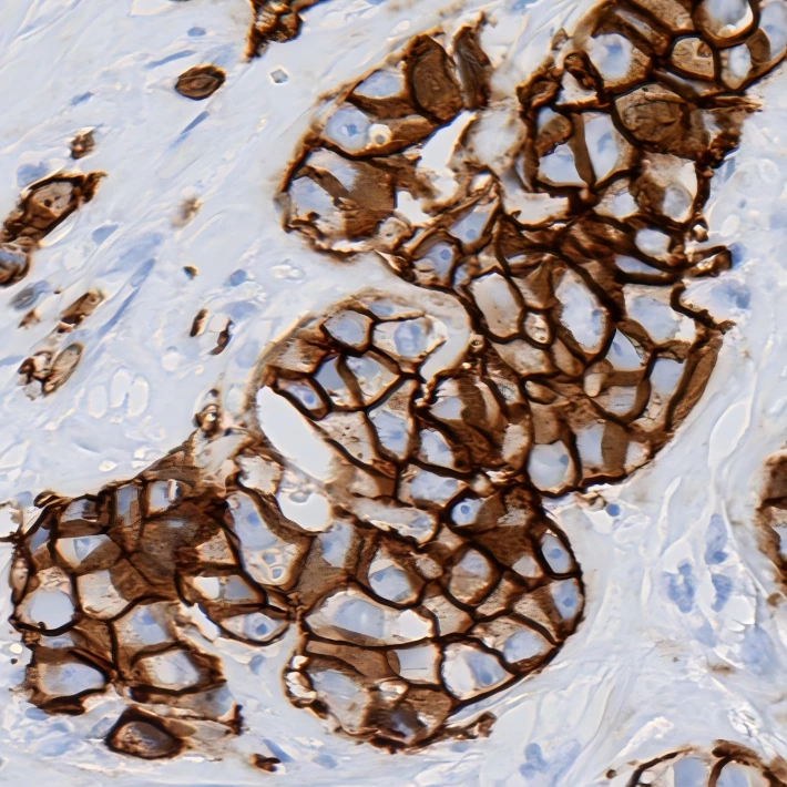
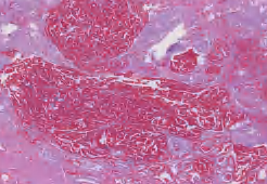
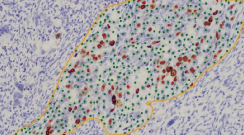
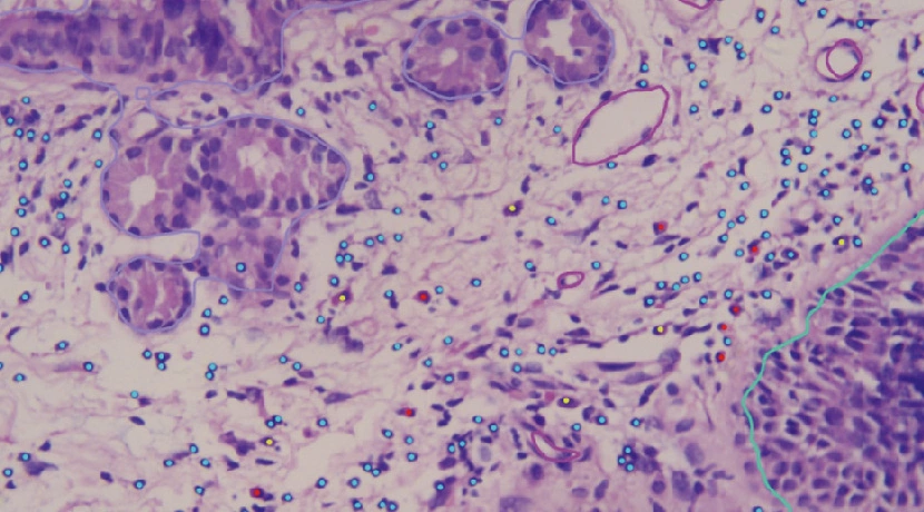
Review IHC nuclear, membrane and cytoplasmic quantification, or nasal-polyp H&E analysis, directly in the live microscopic view. Available with DI-MSA-9.
Digital pathology products for whole-slide scanning, AI analysis, intelligent microscopy, and connected workflows.
Compare scanner models by slide-loading capacity and discuss the configuration that fits your workflow.
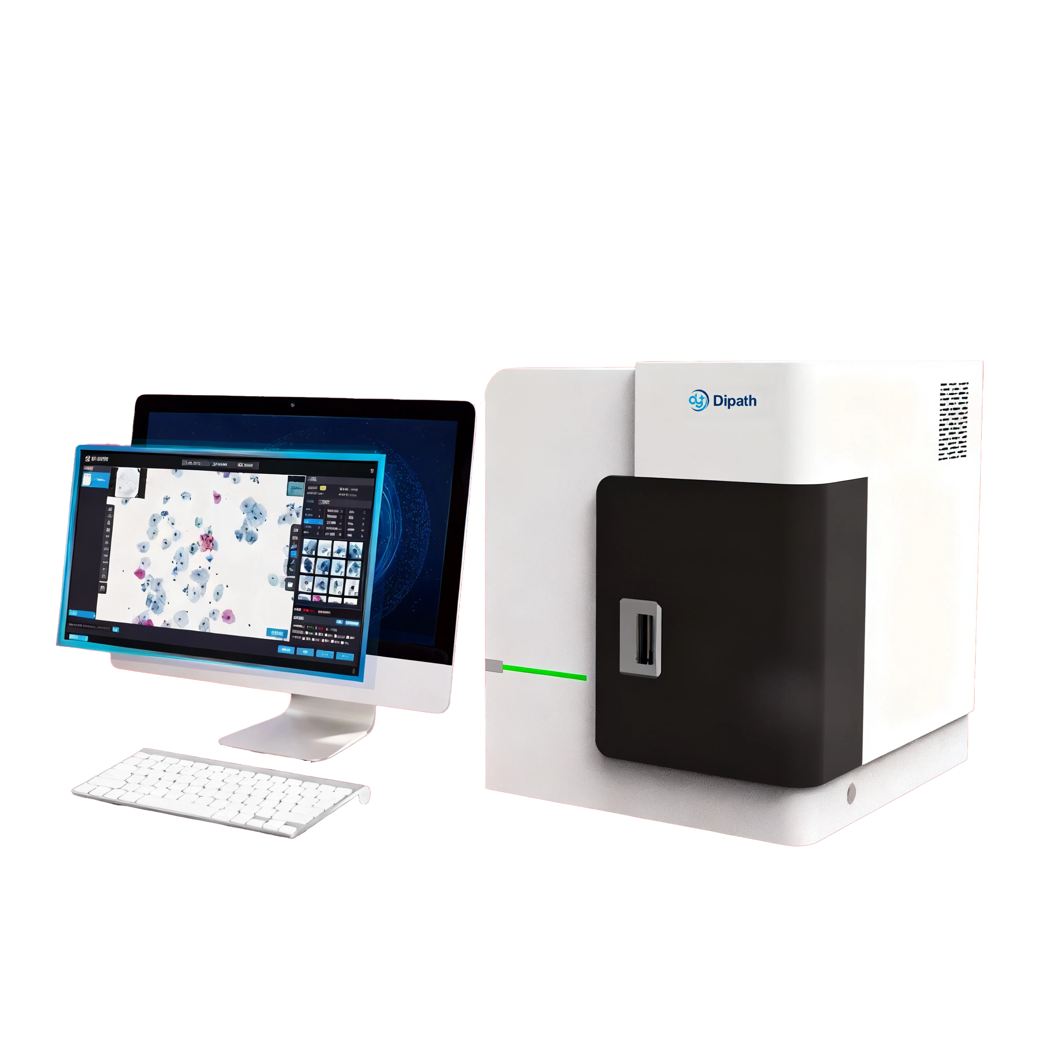
Entry-Level Scanner
A 60-slide configuration for laboratories evaluating or expanding digital slide workflows.
| Slide Capacity | 60 slides |
|---|---|
| Scan time | 20× ≤15 s40× ≤35 s |
| Pixel resolution | 20× ≤0.156 μm/pixel40× ≤0.078 μm/pixel |
| Loading | Loads aluminium-alloy slide holders in a single magazine. |
| Image formats | .csp · .dyj · .tiff · .dcm · .svs |
Scan times shown for a 15 × 15 mm area at the indicated magnification.

High-Capacity Scanner
A 240-slide configuration for workflows with high-capacity batch requirements.
| Slide Capacity | 240 slides |
|---|---|
| Scan time | 20× ≤15 s40× ≤35 s |
| Pixel resolution | 20× ≤0.156 μm/pixel40× ≤0.078 μm/pixel |
| Loading | Loads aluminium-alloy slide holders in dual magazines, with continuous loading. |
| Objective | X-LINE, infinity-corrected |
| Image formats | .csp · .dyj · .tiff · .dcm · .svs |
Scan times shown for a 15 × 15 mm area at the indicated magnification.
The dScan-D200 is a customized dScan-D240 variant available in selected markets. Contact us for configuration options and local availability.
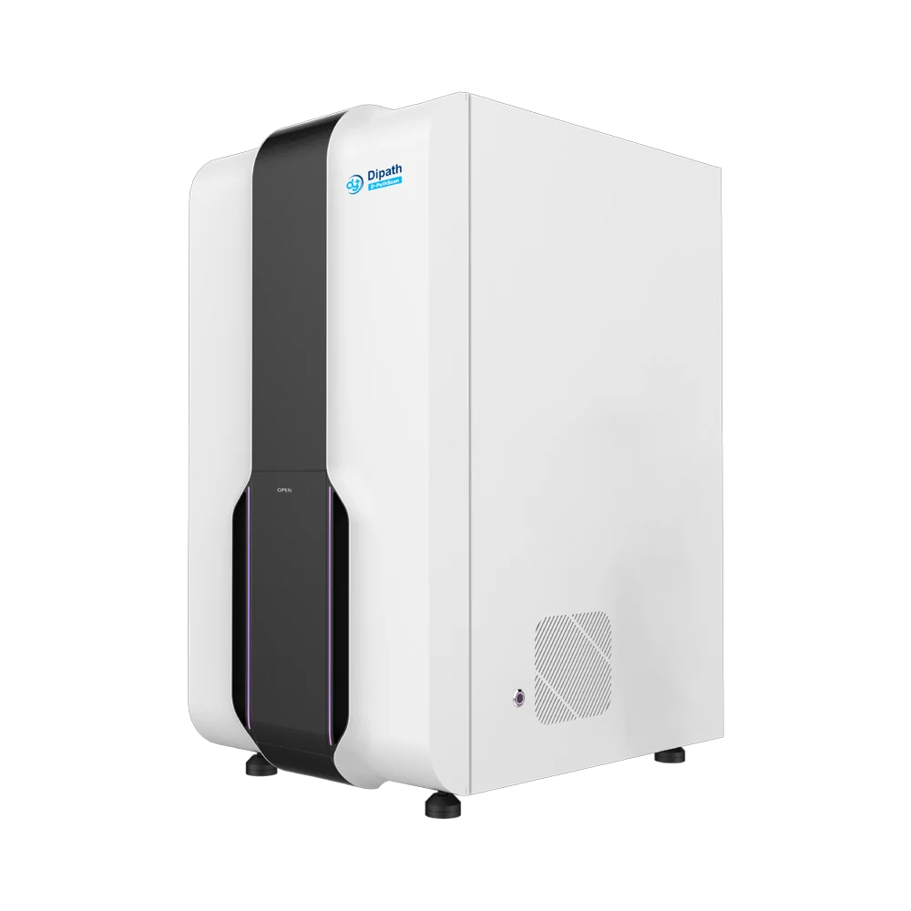
Up to 750 slides
A configuration for larger slide batches, with 15 independent slide racks and capacity for up to 750 slides.
| Slide Capacity | Up to 750 slides |
|---|---|
| Scan time | 20× ≤15 s40× ≤35 s |
| Pixel resolution | 20× ≤0.125 μm/pixel40× ≤0.0625 μm/pixel |
| Loading | Loads 15 independent slide racks, is compatible with multiple types of staining racks, and requires no slide holders. |
| Objective | Two objectives, 20× and 40×; X-LINE, infinity-corrected |
| Image formats | .csp · .dyj · .tiff · .dcm · .svs |
Scan times shown for a 15 × 15 mm area at the indicated magnification.
Objective configurations for 750-slide models: D-PathScan-AI750 (20× and 40×); dScan-750S (40×); D-PathScan-HK (20×).
| Technical specifications | dScan-D60 | dScan-D240 | D-PathScan-AI750 |
|---|---|---|---|
| Slide Capacity | 60 slides | 240 slides | Up to 750 slides |
| Loading | Loads aluminium-alloy slide holders in a single magazine. | Loads aluminium-alloy slide holders in dual magazines, with continuous loading. | Loads 15 independent slide racks, is compatible with multiple types of staining racks, and requires no slide holders. |
| Scan time | 20× ≤15 s40× ≤35 s | 20× ≤15 s40× ≤35 s | 20× ≤15 s40× ≤35 s |
| Pixel resolution | 20× ≤0.156 μm/pixel40× ≤0.078 μm/pixel | 20× ≤0.156 μm/pixel40× ≤0.078 μm/pixel | 20× ≤0.125 μm/pixel40× ≤0.0625 μm/pixel |
| Image formats | .csp · .dyj · .tiff · .dcm · .svs | .csp · .dyj · .tiff · .dcm · .svs | .csp · .dyj · .tiff · .dcm · .svs |
| Workflow fit | A 60-slide configuration for laboratories evaluating or expanding digital slide workflows. | A 240-slide configuration for workflows with high-capacity batch requirements. | A configuration for larger slide batches, with 15 independent slide racks and capacity for up to 750 slides. |
Scan times shown for a 15 × 15 mm area at the indicated magnification.
Objective configurations for 750-slide models: D-PathScan-AI750 (20× and 40×); dScan-750S (40×); D-PathScan-HK (20×).
Open a slide, inspect the image overlays and quantitative results, then review the findings.

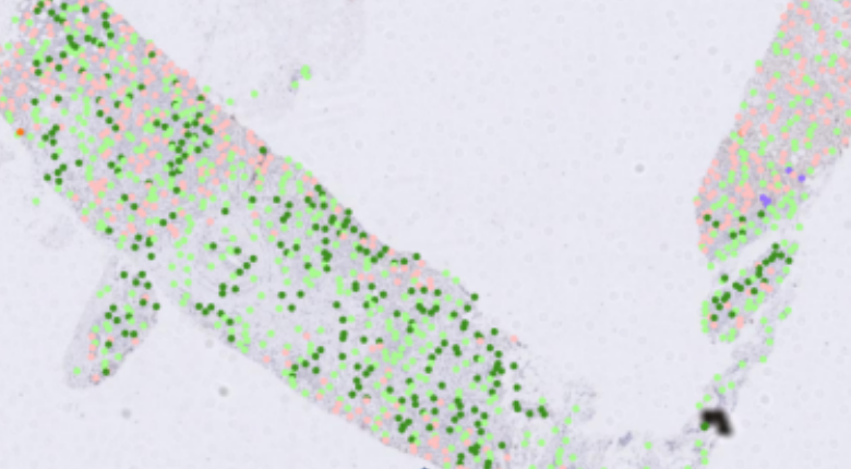
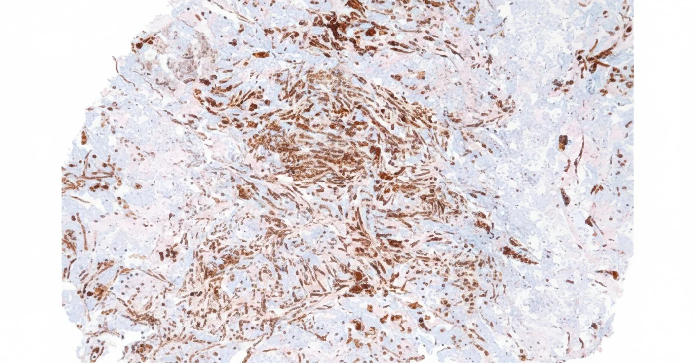
Review cell-level staining overlays and quantitative results for HER2, PD-L1 and Ki-67, with verification by a pathologist.

Locate candidate tissue regions, then inspect the findings in the whole-slide viewer.

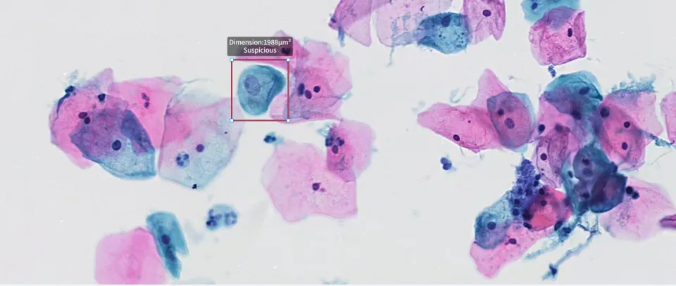
Review candidate cells and their original slide locations.
Biomarker quantification, cytology and tissue analysis.
Identify potential blur, folds, bubbles or contamination.
Navigate from a flagged thumbnail to the affected region.
Use the findings to guide technician review and rescanning decisions.
More applications: ER/PR quantification · FISH spot detection · Research annotation.
Share your assay, staining protocol and deployment needs. We will help confirm the module, software version and market-specific scope.
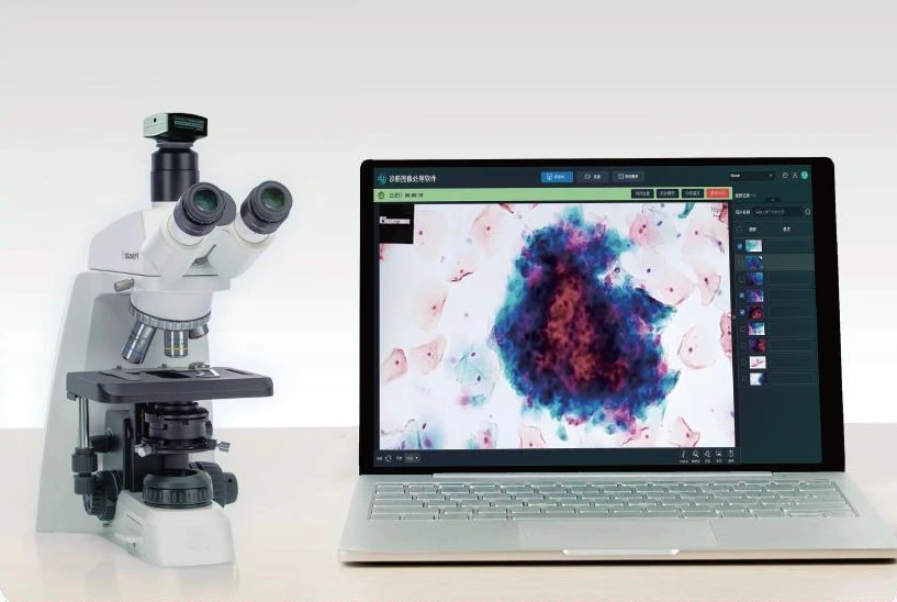
Keep your microscope. Add 4K image capture, live consultation and teaching, with AI analysis and manual whole-slide scanning on DI-MSA-9.
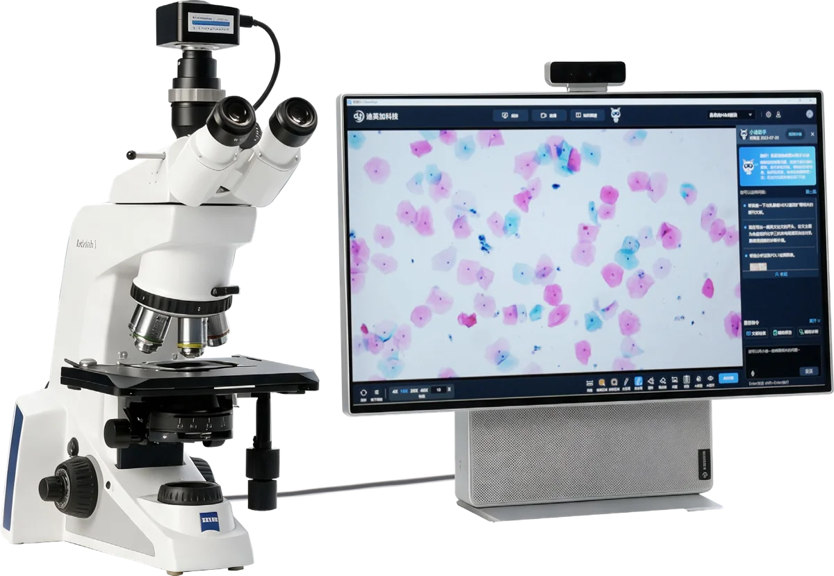
Both models capture microscopic images and support consultation, teaching and annotation. DI-MSA-9 adds real-time AI analysis, manual scanning with image stitching, and DiAssistant.
Powered by DeepPathAI, DiAssistant supports literature and knowledge retrieval, voice interaction, pathology content tasks and report drafts for pathologist review. Available with DI-MSA-9.
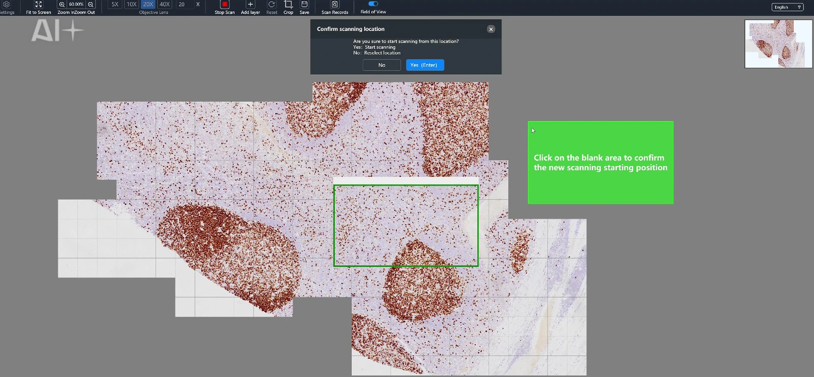
Move the slide by hand while D-CleverEye stitches the captured fields in real time. Scan a selected region or extend the capture across the whole slide.
Available with DI-MSA-9.
Review IHC nuclear, membrane and cytoplasmic quantification, or nasal-polyp H&E analysis, directly in the live microscopic view. Available with DI-MSA-9.
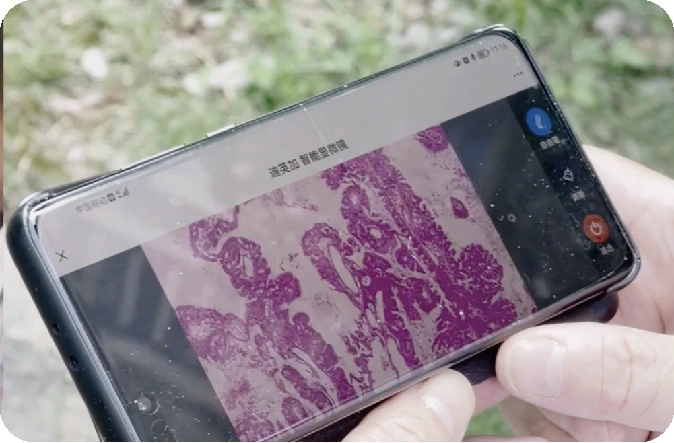
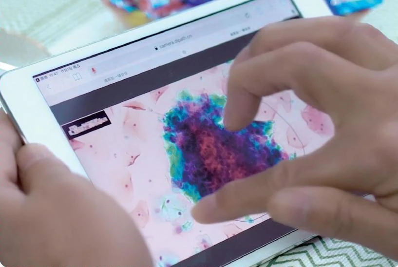
Share a link or QR code to review the same live view, annotations and voice across desktop and mobile devices. Supports remote consultation, intraoperative frozen-section consultation and teaching.
| Component or capability | DI-MSA-9 | DI-MSB-8.3 |
|---|---|---|
| Camera | 9 MP CMOS | 8.3 MP CMOS |
| Workstation | 4K display + PC | All-in-one PC |
| Image AcquisitionLive Consultation5G TransmissionLive TeachingImage Annotation | Supported | Supported |
| DiAssistant | Supported | Not included |
| AI Quantification | Supported | Not included |
| Knowledge Graph | Supported | Not included |
| Manual WSI scanning and real-time stitching | Supported | Not included |
| Core Expert Network | Supported | Not included |
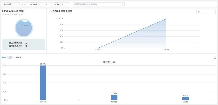
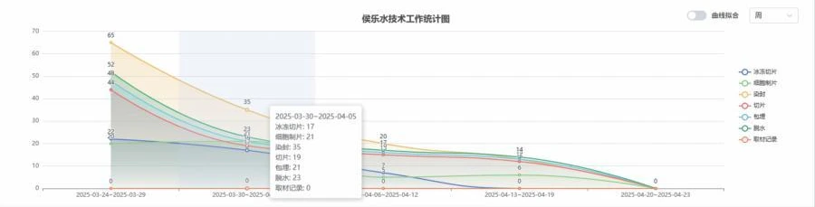
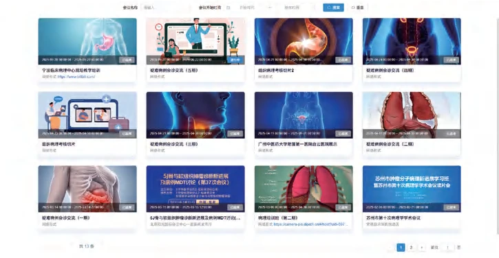
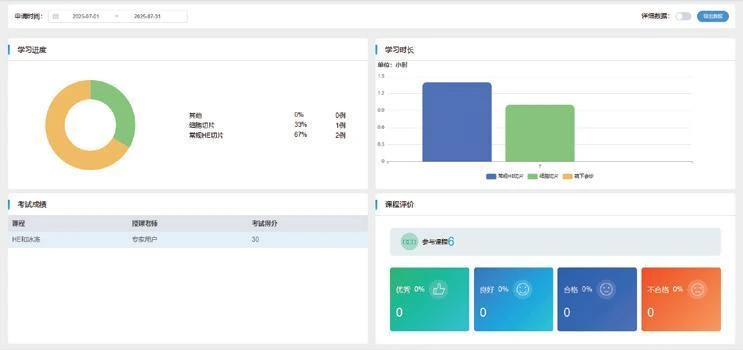
Tools for slide management, consultation, laboratory information and teaching.
Pathology foundation model
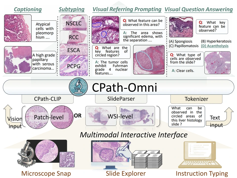
A multimodal pathology model brings microscope fields and whole-slide images into one framework.
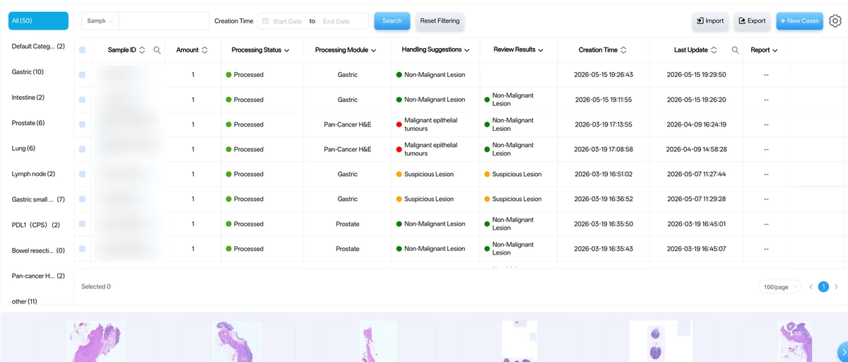
Digital slide management
Keep images, review tasks and controlled sharing together.
Remote pathology consultation
Share selected slides or live images for expert discussion.
Pathology quality control and information management
Link specimen records, processing and report review.
Pathology training and teaching
Bring teaching slides, discussion and assessment together.
Capabilities shown are high-level workflow descriptions. Availability, configuration, intended use, and regulatory status vary by model, version, and market. Contact us for a scoped review.