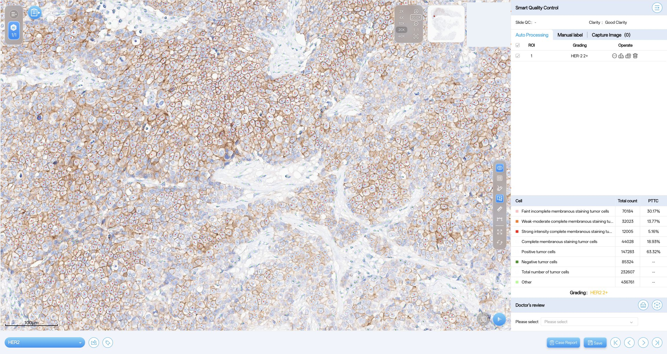
Review membrane-staining annotations, tumor-cell counts and quantitative findings alongside the original HER2 IHC image. A pathologist reviews the outputs and confirms the interpretation.
Breast HER2 IHC whole-slide images. The documented workflow excludes ductal carcinoma in situ (DCIS).
Outputs
Cell-level membrane-staining annotations, cell counts and percentages of total tumor cells (PTTC), grouped by the staining pattern.
Review
Review the annotations and quantitative findings in the source tissue, using the laboratory’s antibody clone, staining protocol and HER2 scoring scheme.
Supported analysis and intended use depend on the module, software version and market. Product example findings and clinical-study results describe different contexts and should be read within those contexts.
A defined analysed-region example
Original product example at 20.0×. Findings refer to the analysed region and require pathologist review.
HER2 2+ example — analysed region
Finding
Cell count
PTTC
Total tumor cells
232,607
—
Positive tumor cells
147,283
63.32%
Faint, incomplete membrane staining
70,184
30.17%
Weak–moderate, complete membrane staining
32,023
13.77%
Strong, complete membrane staining
12,005
5.16%
Negative tumor cells
85,324
—
Other cells
436,761
—
PTTC is the percentage of total tumor cells. Only selected staining categories visible in the source software are listed; they are not a complete breakdown of all positive cells. Browsing image fields does not run a new analysis or change the original region findings.
Wu et al. · Signal Transduction and Targeted Therapy 11, 241 (2026)
The study combined pretreatment H&E and HER2 IHC biopsy images with clinical variables to study response to neoadjuvant dual HER2-targeted therapy in HER2-positive breast cancer. Model development used 276 cases, with temporal (82), trial-based (85) and external (40) validation cohorts.
Software role
A customized D-PathAI pipeline measured membrane-staining intensity and completeness. HoVer-Net analysed H&E images, and a separate research framework combined the features.
Limits
Retrospective analyses can introduce selection bias. Prospective validation and staining/scanning standardization are needed before wider clinical use.
HER2 scoring: reader agreement and remaining limits
Dong et al. · npj Breast Cancer 12, 9 (2026)
The study included 1,455 retrospective cases, a 45-laboratory staining assessment and a separate 209-slide comparison of manual and AI-assisted readings. These components address different questions and are not one combined accuracy cohort.
Software role
D-PathAI assisted HER2 IHC interpretation in the 209-slide comparison. AI-assisted readings showed higher agreement within that dataset.
Limits
Cell-detection errors and nonspecific staining persisted. Ultralow/null separation remained unreliable, and manual verification was required.
These studies describe their published workflows. D-PathAI was used for image analysis; these studies do not establish treatment-selection functionality in the current product. Neither the region example nor the study summaries establish performance for every current module, scanner or deployment.
Prepare a module evaluation
Specify the breast-cancer use case, antibody clone, staining protocol and required HER2 scoring scheme.
Provide representative images, scanner and file details, and the review procedure used by the laboratory.
Agree the module version, analysis-region definition, output checks and pathologist review before deployment.