DeepPathAI
A multimodal foundation for pathology image analysis, professional Q&A and workflow applications.
PATHOLOGY FOUNDATION MODEL
From image understanding to everyday pathology
DeepPathAI brings pathology images, case information and professional knowledge together. It works with D-PathAI and D-PathPIS to support specialist analysis, pathology Q&A and reporting assistance, and provides a foundation for joint development with hospitals and industry partners.
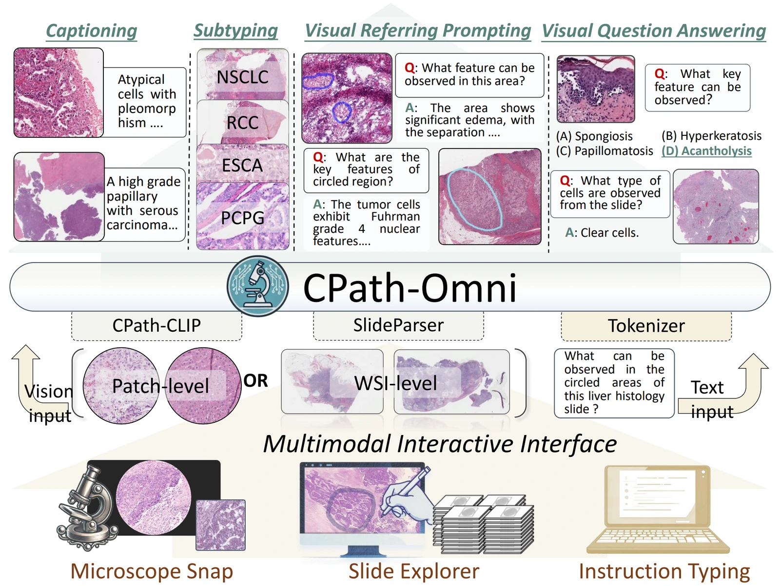
CPath-Omni CVPR 2025
One framework for image patches and whole slides
CPath-Omni brings classification, image description, visual Q&A and region-specific prompting into a unified research framework. These capabilities underpin the development of flexible pathology applications across image scales.
- Visual representations
- Learn tissue architecture and cell morphology to support specialist classification, retrieval and image-analysis development.
- Image–text alignment
- Connect pathology images with professional descriptions, providing a foundation for image–text retrieval and multimodal application development.
- Multimodal interaction
- Combine images, case context and text instructions for image Q&A, information synthesis and reporting assistance.
PathMMU ECCV 2024
Evaluate understanding with pathology expertise
PathMMU uses image-based questions and answer explanations, with validation and test questions reviewed by pathologists. It examines professional understanding, reasoning and robustness to image changes, providing a reference for model evaluation and specialty adaptation.
View the original paper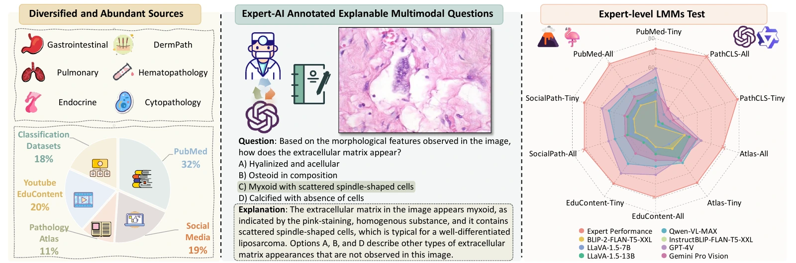
PathMMU is a research benchmark. Its scores do not establish clinical diagnostic performance.
D-PathPIS · MODEL IN THE WORKFLOW
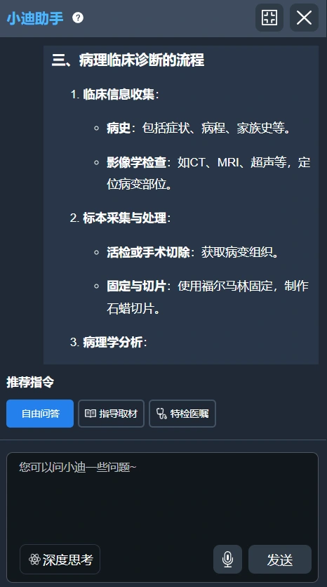
Pathology assistance within the case workflow
Bring case information and a preliminary diagnosis into pathology Q&A, report drafting and content review. The pathologist reviews the suggestions and confirms the final report within the existing workflow.
- Case context
- AI assistance
- Pathologist confirmation
DeepPathAI
Functions are configured for the specialty, software version and project.
HOSPITALS AND INDUSTRY PARTNERS
Build around a defined specialty need
Develop a specialty model or integrate AI into an existing product. We work with partners on model adaptation, local knowledge and workflow integration, with a jointly agreed evaluation and rollout scope.
Deployment and integrationDefine the task
Agree the specialty use case, available data and evaluation scope.
Adapt the model
Adapt to specialty images, local knowledge and reporting conventions.
Connect applications
Integrate with agreed image-review, reporting or quality workflows.
Evaluate together
Review with pathologists and confirm the deployment scope.
Explore AI-assisted slide review
Explore the product screen, then select a local field to inspect the image and analysis findings.
 D-PathAIProduct examples
Loading product image…This source field could not be loaded. Choose another field or analysis example.
Product screenshots and original image fields. Analysis findings require a pathologist’s review.
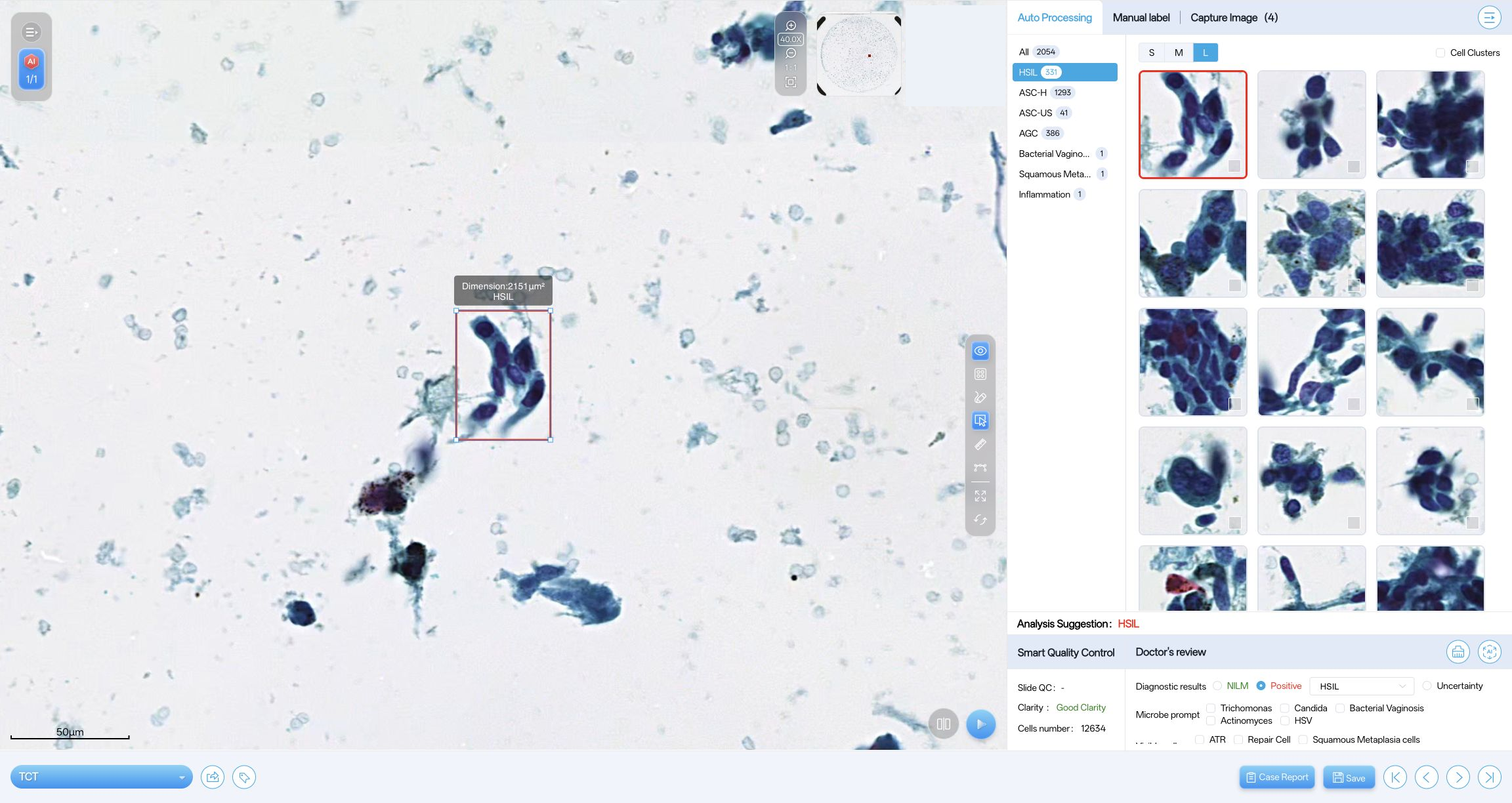
Cervical cytology
Review the product overview and original cell fields with AI candidate markers.
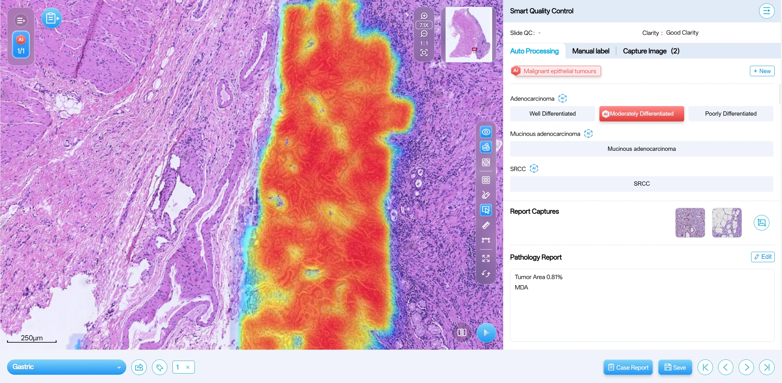
Stomach H&E
Review the tissue heatmap and source classification in the product overview.
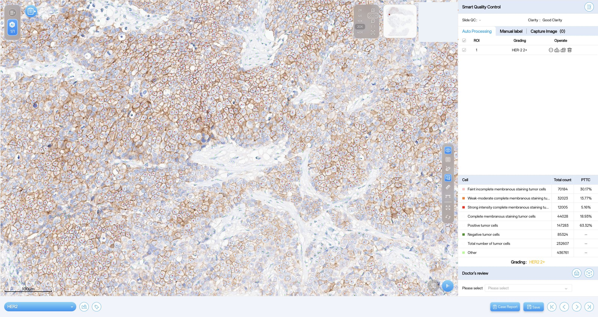
HER2 IHC
Inspect membrane-staining overlays and cell counts for the analysed region in this product example.
Supported analysis and intended use depend on the module, version and market. Outputs require review by a qualified user.
Plan deployment with your laboratory and IT team
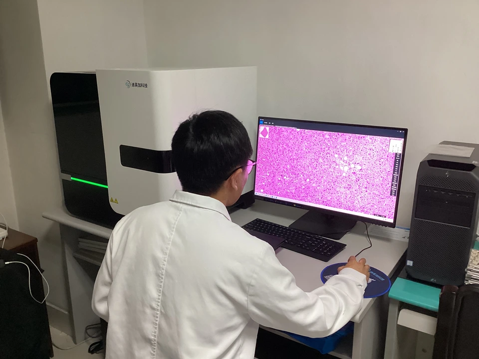
Start with your scanner, image formats, LIS and data-handling requirements. The final configuration depends on the agreed product and deployment.
- On-premises deployment
- Review server and storage requirements, user access, software updates and responsibilities with your IT team.
- Cloud deployment
- Confirm the hosting location, data access, retention, backup and contractual arrangements for your project.
- Scanner and LIS integration
- Share scanner models, file formats and LIS interfaces so the team can assess compatibility and the work needed.